The CICL tailors our full service offerings to best meet the specific needs of each study.
In addition to providing high-quality quantitative and qualitative imaging analysis, the CICL offers complete on-site and off-site training including live demonstrations, site instruction manuals, and site certification. The CICL works closely with Sponsors to define the most appropriate Scope of Work for each project. Specific image processing and analytic services include comprehensive analysis of:
- Echocardiograms (echo)
- Cardiac computed tomography (CT)
- Cardiac magnetic resonance imaging (MRI)
- Arterial tonometry
Echocardiographic Analysis
Comprehensive quantitative analysis of echocardiograms include:
- Assessment of left ventricular size (areas, volumes, ejection fraction)
- Assessment of right ventricular size (areas)
- Assessment of left ventricular function (ejection fraction)
- Assessment of right ventricular function (fractional area change)
- Quantitative assessment of regional wall motion
- Assessment of ventricular wall thickness
- Quantitative assessment of mitral regurgitation
- Comprehensive Doppler analyses
- Comprehensive LV remodeling analyses
- Strain and dysynchrony analyses by speckle tracking (vendor independent)
- 3D comprehensive assessment of cardiac structure and function (vendor independent)
- 3D strain and dysynchrony analyses by speckle tracking (vendor independent)
- 3D analyses of torsion by speckle tracking (vendor independent)

Cardiac CT Analysis
Comprehensive quantitative and qualitative analyses of cardiac CT include:
- Coronary artery/bypass graft assessment: reference luminal diameter, minimal luminal diameter, reference luminal area, and minimal luminal area
- Coronary angiography assessment
- Coronary artery calcium quantification
- CT assessment of LV volumes and ejection fraction

Cardiac MRI Analysis
Comprehensive quantitative analyses of cardiac MRI include:
- Assessment of ventricular global size and function
- Assessment of ventricular mass
- Assessment of ventricular size (areas, volumes)
- Quantitative regional function (with tagging and other quantitative techniques)
- Quantitative valvular regurgitation fraction and regurgitant volume
- Quantitative assessment of valvular stenosis
- Myocardial infarct mass, infarct size, and transmural infarct extent
- Myocardial infarction tissue characterization (peri-infarct region, microvascular obstruction)
- 2D and 3D great vessel structure and anatomy, lumenal blood flow, and shunt ratio
- 3D-Pulmonary venous anatomy
- Myocardial and liver iron content

Vascular Tonometry Analysis
Comprehensive quantitative analyses of arterial pulse waveforms include:
- Average +/- standard deviation of augmentation index
- Pulse wave velocity
- Average late systolic peak pressure
- Brachial and radial artery pressures
- Early and late systolic radial artery pressures

Workflow
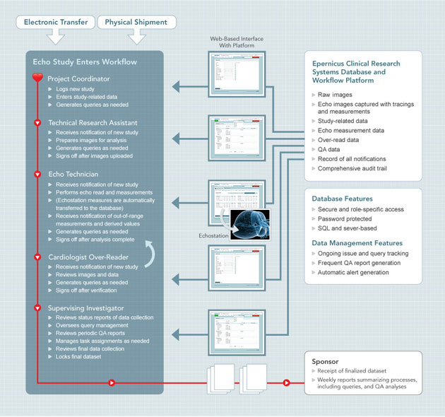
The CICL workflow is supported by a robust, customized Clinical Research Systems (CRS) platform that provides a seamless database-driven system for study tracking, query generation, capture of image analysis data, management of study-related data, and management of study workflow.
Front-end interfaces to the CRS platform reside on the workstations of all research staff involved in the project, including project coordinators, technicians, physician over-readers, and investigators. Access to the CRS platform is password-protected for use only by authorized personnel. In addition, access to data entry, review, edit, and management features is role-specific (i.e. allowing selective access to administrative, technical, and over-read options, depending on the user). All image acquisition and image analysis data that is captured and managed by the CRS platform is housed in a secure, industrial strength SQL relational database system that includes robust data replication and backup systems.
The CRS platform allows work to be performed by multiple members of the research team in parallel rather than in series. Compared to conventional paper-based or multisystem-based methods for managing a large amount of complex research data across multiple sites, the CRS platform allows for shared image data capture, storage, analysis, and management to occur in a high quality and time efficient manner while minimizing errors.

